Welcome
Changing minds through the power of the smile.
Improving the brain-body connection and airway function through optimizing the oral environment
Improving the brain-body connection and airway function through optimizing the oral environment
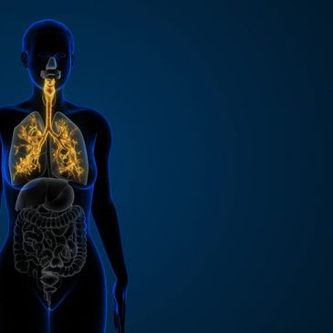


The neural circuits in our bodies are equivalent to the electrical wiring systems in our homes.
They carry the signals and energy that make things "work".
The neural circuits of the mouth that control some of the most important and intimate aspects of our lives are influenced by the tongue, lips, teeth and bones that dentists are uniquely trained to understand and treat.
However, traditional dental training doesn't explore the many aspects of human physiology that the emerging science is connecting directly to the role of the dental provider in influencing human health and function.
This influence begins before we are born and doesn't end until we take our last breath.
The concepts related to Neurodevelopmental Dentistry promote an enlightened awareness of the many exciting and influential aspects of dental and orthodontic care as related to everything from infant feeding to neurodenerative conditions of the elderly population.

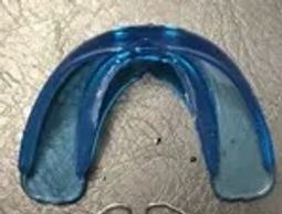
There are many options available to "open the airway" during sleep that pull the lower jaw (mandible) forward to reduce the effects associated with snoring.
What if the same device can be used to improve the brain body connection for improved treatment outcomes?
Regardless of the age or capabilities of a person, the principles of neurodevelopmental wellness are the same.
Dental devices that are designed to improve intellectual and athletic performance are based on principles and assessments that improve both functional capacity and quality of life.
Devices designed to improve the function of the swallow and those improperly used as compensations for a dysfunctional swallow are often an integral part of a comprehensive approach topatient care.
Some dental devices are specifically designed to compliment the sophistication of the cranial complex and its contents. (the brain)
Others are designed to simply create structural changes.
Combining the appropriate dental therapies with careful patient selection is critical to successful treatment outcomes.

Dental devices that are intentionally designed to guide proper tongue motions and placement provide the gift of continuity to a treatment approach that emphasizes efficiency and successful treatment outcomes. By avoiding the frustration and inconsistent outcomes of repetitive exercises without a thourough understanding of the neuroogical aspects of the swallow, patients often appreciate the cost and time savings of the use of neurofunctional dental devices.

The Synergy Academy offers AGD PACE approved coursework on the subjects of ALF Therapy, OraLase, BabyLase, RestOralase and Neurodevelopmental Dentistry.

Helping our most vulnerable to become our most victorious through the gentle miracle of light.
Non-surgical laser-based therapy to improve infant feeding by releasing tongue restrictions and improving the brain-body connections that can be disrupted during pregnancy, childbirth and the first 2 years of life.

An innovative and comprehensive approach to addressing craniofacial pain, dysfunctional neural circuits, fascial restrictions and sleep dysfunction through the utilization of dual laser wavelengths (NdYAG & ErYAG) in conjunction with neuromodulatory dental appliances.

Non-surgical correction of dysfunctional neural circuits as well as tongue ties and fascial restrictions.

SImply sophisticated dental devices designed to the optimize the power of the mental-dental connection to simultaneously improve form and function.
The SWITCH Assessment is an acronym for Strength With Integration of Trigeminal and Cranial Harmony. Neurological signals originating from the swallow and the bite affect the postural control of the body's musculoskeletal system through the locus coeruleus and vestibulospinal pathways.
Those are a lot of big words to describe a simple concept: The body and brain are either connected...or disconnected through the neural circuits of the mouth.
The SWITCH Assessment has been designed to not only educate the on the level of function and resilience of the patient, but also to guide treatment sequencing and decisions.
Subjective Test Of Pain
Pain upon palpation often indicates a dysfunctional movement pattern which should be a consideration in assessing a patient for efficient chewing and swallowing.
The Starfish Assessment provides valuable information related to the presence (retention or return) or a primptive reflex known as the Moro Reflex, which is often referred to as the startle reflex.
If this primitive movement pattern is being inappropriately expressed, the fascial system of the body often presents as tight or restrictive. Additional symptoms include: (but are not limited to)
Identifying the presence of an active Moro reflex in a child or an adult can make a tremendous difference in successful treatment outcomes.
Successful treatments result in quality of life improvements as well as relieving the fascial restrictions that can be misdiagnosed as a tongue tie.

"Based on these findings, we conclude that a dysfunctional mesencephalic TRIGEMINAL nucleus (MTN) may be incapable of activating neurons in the ascending reticular activating system (ARAS),..., and that this will ultimately lead to sleep apnea."
Despite its name, obstructive sleep apnea (OSA) is not caused by an airway obstruction that prevents breathing.
Andrisani, G., Andrisani, G. Sleep apnea pathophysiology.
Sleep Breath (2023).
In other words, we do not clench our teeth to support our airway as previously thought. The science shows that we clench our teeth until we deplete the neurotransmitters that allow us to continue to breath while sleeping....and then sleep apnea occurs.
Teeth clenching is often a compensation for a dysfunctional swallowing pattern, which then evolves into continued dysfunction through the creation of dental diseases and whole-body compensations
The research shows that the floor of the mouth is composed of fascia that is connected to the entire body. It also shows that if the muscle under the tongue (genioglossus) has been damaged during the surgical procedure to remove the tongue tie, that another restriction has been created.
This might explain why some people experience temporary benefits of a tongue tie release (honeymoon) while others receive the beneit of a long-lasting improvements (marriage). Regretfully, some unfortunate patients receive either no benefit or are worse after the procedure.
The research on treatment success is often inconclusive. Patient feedback is exceptionally variable. Professional politics are uncomfortable.
Could the reason for such confusion be related to the unaddressed, yet impactful consequences of dysfunctional neural circuits and fascial restrictions that we not addressed prior to the surgical release?
The lingual frenulum is not composed of connective tissue fibers that have an anteroposterior orientation nor is it a discrete cord or band as often described in literature (Hong et al., 2010; Ghaheri,2014; Watson-Genna, 2017).
In contrast, the fascial fibers that form the frenulum have a basket-weave orientation as they cross the midline.
Furthermore, the diagnosis and classification of a “posterior tongue tie” is based on the construct of a midline “submucosal band” and is notsupported by these dissections.
Mills, N., Pransky, S. M., Geddes, D. T., & Mirjalili, S. A. (2019).
What is a tongue tie? Defining the anatomy of the in-situ lingual frenulum. Clinical Anatomy
The histochemical structure of the deep fascia and its structural response to surgery
J Hand Surg Br. 2001 Apr;26(2):89-97.
McCombeD1, Brown T, SlavinJ, Morrison WA.
"This structure was also evaluated after it had been raised as a fascial flap and in another site after the underlying muscle surface had been disrupted.... The post-surgical specimens demonstrated preservation of the structure of the interface between fascia and muscle, including the retention of the hyaluronic acid lining, if the epimysium was intact.
However, if the epimysium was disrupted, the structure of the interface was obliterated.
https://pubmed.ncbi.nlm.nih.gov/11281657
Signaling to the brainstem through the trigeminal nerve as a result of balanced forces of the swallow and bite create the functional neural circuits that lead to improved brain and body function
Neurodevelopmental treatment goals include an acute awareness of assessing and maintaining balanced neural circuits and functional movement patterns to prevent the development of the dysfunctional neural circuits that lead to sleep apnea and other neurocognitive conditions.
It is known that asymmetries in trigeminal (as well as vestibular) signals induced by malocclusion are detrimental for performance and their elimination by occlusal correction improves cognitive performance in patients affected by AD as well as in normal subjects.
Trigeminal, Visceral and Vestibular Inputs May Improve Cognitive Functions by Acting through the Locus Coeruleus and the Ascending Reticular Activating System: A New Hypothesis
https://www.frontiersin.org/articles/10.3389/fnana.2017.00130/full